 My Cart
 Measuring Melanin and Hemoglobin levels of Skin Samples
Background/Applications:
Human skin measurements apply to many industries and field studies with differing analyses in absorbance, transmission, and or reflectance. In this experiment, we tested the absorbance and reflectance of skin samples to analyze skin pigmentation and protein structures.
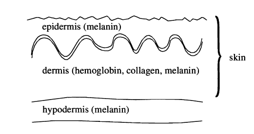
Figure 1: Layers of the skin
The skin pigmentation studied in this experiment was melanin. Melanin is mainly located in the epidermis (Figure 1) and is responsible for the pigmentation of the skin, hair, and eye color. The more melanin an individual produces, the darker their eyes, hair, and skin will be. The amount of melanin in a body depends on a few different factors including genetics and how much sun exposure their ancestral population had.
The protein studied in this experiment was hemoglobin. Hemoglobin is located in the dermis which is below the epidermis. Oxygenated hemoglobin has absorption bands at 420 nm and in the 545- 575 nm range. The latter range produces an M-shaped pattern in absorbance readings. These absorption bands occur when hemoglobin is bound with oxygen. Heavily pigmented skin has increased amounts of melanin which absorbs most of the light in the epidermis, allowing a much smaller percentage of incident light to reach the dermis. Because of this, the hemoglobin peaks are more sensitive to detect spectroscopically than the melanin peaks when an individual gas darker skin.
The skin reflectance measurements were used to show what colors were reflected from the skin samples and at what intensity. The more light the skin absorbs, the less light the skin will reflect. So, for an individual with a high amount of melanin, the melanin will absorb most of the light causing the reflectance to be lower.
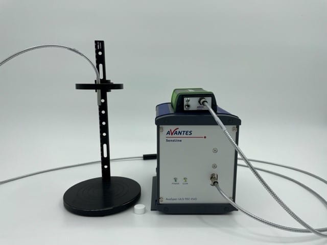
Figure 2: System Used for Experiment
Description of System:
For this experiment, we used Avantes’ top-of-the-line UV/VIS spectrometer, the AvaSpec-HERO (Figure 2). The AvaSpec-HERO is built up around our High-Sensitivity, Compact (HSC), 100mm optical bench offering a numerical aperture (NA) of 0.13 and a cooled, back-thinned CCD detector (1024×58 pixels). Electronics-wise, the HERO uses our state-of-the-art AS7010 board, which includes a high-performance Analog Digital converter with excellent noise performance and the ability to support high-speed communication through USB 3.0 and or gigabit ethernet. Due to the high sensitivity of this instrument, the HERO can help to reach the dermis to read the hemoglobin measurements.
The light source used was the AvaLight-HAL-S-Mini which is a Tungsten-Halogen light source that is commonly used for spectral measurements in the visible to near-infrared wavelength ranges. It’s a compact, stabilized halogen light source, with adjustable focusing of the fiber connection, maximizing output power at the desired wavelength. The light source also has adjustable output power to provide extra power or longer lamp life depending on the application needs. This light source was chosen for the experiment because the AvaLight-HAL is ideal for absorption/transmittance measurements in the visible spectrum. The wavelength range of this light source is 360- 2500 nm. The AvaLight-HAL-S-Mini also features an internal TTL-shutter, controllable from an AvaSpec Spectrometer. This gives the ability to periodically shutter the light source to take a dark during an experiment or process. This shuttering can be done manually or via software control.
The fiber component used in this setup was Avantes’ FCR-7UVIR400-2-ME reflectance probe. It is a bifurcated cable that uses light from the light source through its six illumination fibers in one leg which converge into the common cable where a single 7th fiber in the center of the light source fibers reads the reflected light at the probe tip. The 7th fiber is coupled to the spectrometer configured to the appropriate wavelength range of interest. Reflectance probes are ideal for skin measurements since it offers direct contact to the sample.
The last component used in this experiment was the WS-2 reference tile. This was used to take a light reference before any skin measurements were taken. The WS-2 is made out of diffuse PTFE material which offers long-term stability, even in UV applications.
Description of Methodology:
During an Avantes open house event, thirty volunteers offered to be a part of this experiment (Figure 3). The volunteers varied in age, gender, and ethnicity. Before testing, an area of the underside of the forearm was chosen and wiped down with an alcohol wipe. The underside of the forearm was chosen for testing so that arm hair and freckles/moles were not in the sample. The underside of the forearm is also hidden away from external factors that can periodically change the color of the skin like the sun. The alcohol wipe was used to remove any unwanted dirt, lotion, and or other products from the skin to provide a clean testing spot. We also made sure that the testing spot was not over a vein.
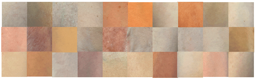
Figure 3: Skin samples of individuals included in this experiment
A WS-2 reference tile was used to auto-configure the software to have an integration time of 10.5 µs and an average of 65. This tile was also used to take a light reference before skin samples were tested. A new light reference was taken between every couple of volunteers or if there were a large time span in-between volunteers. The reflectance probe was also cleaned with an alcohol wipe before every new light reference.
After a suitable testing spot was prepped and cleaned, the volunteers then put their skin underneath the reflectance probe. The testing spot made contact with the skin, but it was not deeply pressed into the probe. The spectra were then saved in absorbance mode after a couple of seconds of software reading. Along with the spectral reading, each volunteer’s eye and natural hair color was documented. Out of the thirty volunteers, ten had dark hair and dark-colored eyes, twelve had dark-colored eyes and light-colored eyes, and eight had light hair and light-colored eyes; with dark-colored eyes meaning brown, and light-colored eyes meaning green, blue, or hazel. Natural light hair and dark-colored eyes is a rare combination and none of our participants fell under that category so it was not included in the analysis.
Test Data and Results:
All thirty volunteers’ skin absorbance spectra can be seen in Figure 4. Absorbance units are unitless (A.U.) because they are calculated from a ratio. The ratio is the intensity of light transmitted through the sample to the light transmitted through a blank (the reference tile). The absorbance graph can show the melanin and hemoglobin peaks in the skin samples. The first major peaks around 370 nm are the melanin peaks. The peaks are quite broad after they hit their max height. The highest melanin sample from a volunteer had an absorbance of about 1.25 A.U., and the lowest melanin sample had an absorbance of 0.36 A.U. The melanin peaks then run into the first hemoglobin peak at around 420 nm which is a singular hump. The second hemoglobin wavelength range is 545-575 nm. The hemoglobin peaks can be seen in that range with some of the samples providing the M-shaped pattern.
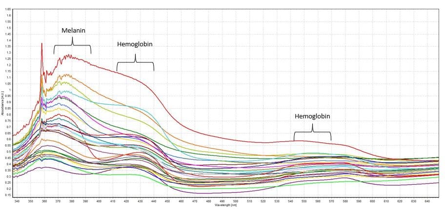
Figure 4: Skin absorbance spectra
The next figure shows the same data in a reflectance graph. Reflectance is measured in percentage going up to 100%. The reflectance graph is relatively the inverse of the absorbance graph. The more light absorbed, the less light can be reflected, meaning that the lowest melanin sample has the highest reflectance. The lowest melanin sample peaks had a reflectance percentage of 68% at 470 nm (blue) and of 59% at 610nm (orangish-red). The highest melanin had a reflectance percentage of 22% at 470 nm and of 36% at 610nm.
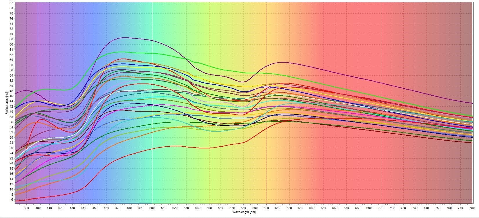
Figure 5: Skin reflectance spectra
Analysis:
The highest melanin samples all belonged to the volunteers with dark hair and dark-colored eyes (Figure 6). The absorbance range for the melanin peaks was from 0.66 A.U. to 1.25 A.U. The first hemoglobin peak at 420 nm varied between all samples, but they all had a broad hump-like shape. The second hemoglobin peak range from 545-575 nm did not deliver any distinct M-shape. This was expected since high amounts of melanin absorbs most of the light in the epidermis which does not allow a lot of light through the dermis where the hemoglobin is.
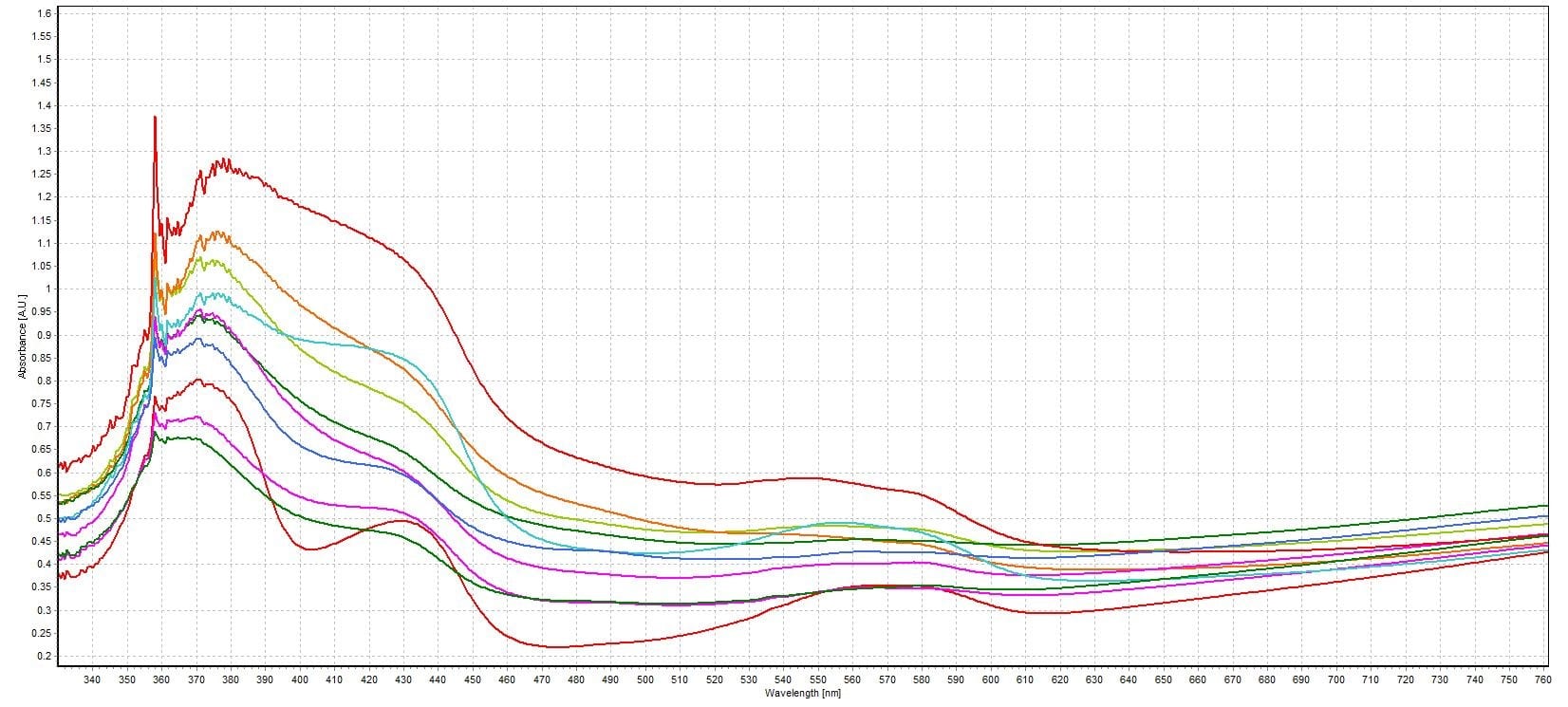
Figure 6: Volunteers with dark hair and dark-colored eyes
The largest number of volunteers were in the dark hair and light-colored eyes category (Figure 7). The absorbance range for the melanin peaks was from 0.44 A.U. to 0.86 A.U. The first hemoglobin peaks at 420 nm had a more defined shape that was more uniform between samples, and the second hemoglobin peak range from 545-575 nm started to develop more of an M-shape as well.
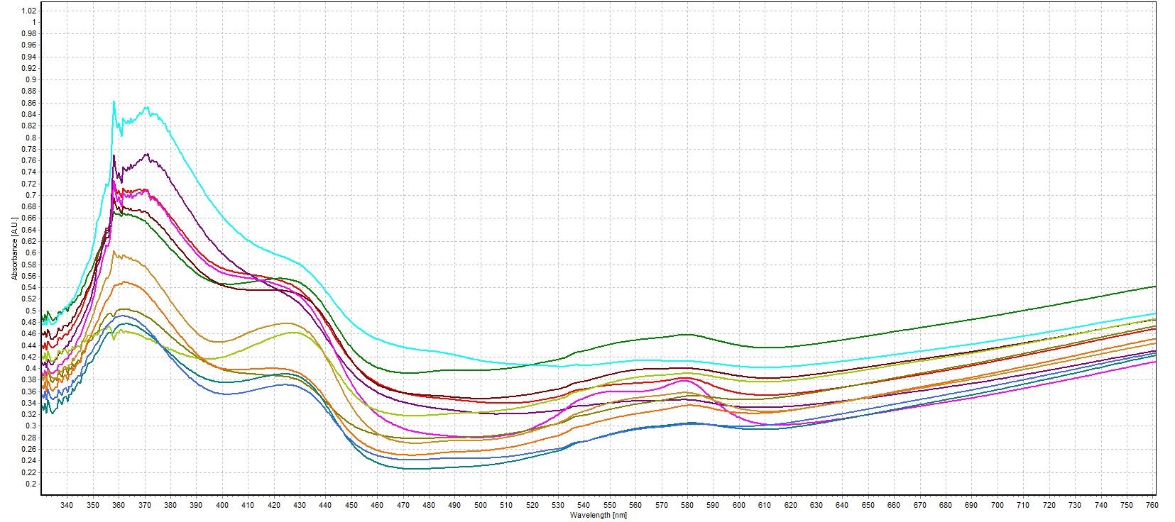
Figure 7: Volunteers with dark hair and light-colored eyes
The lowest melanin samples all belonged to the volunteers with light hair and light-colored eyes (Figure 8). The absorbance range for the melanin peaks was from 0.36 A.U. to 0.73 A.U. The first hemoglobin peaks at 420 nm had the most defined shapes that were fairly uniform between samples, and the second hemoglobin peak range from 545-575 nm finally developed the M-shape hemoglobin peaks. The lower amounts of melanin allowed light to reach the dermis where the instrumentation could then detect more of the hemoglobin.
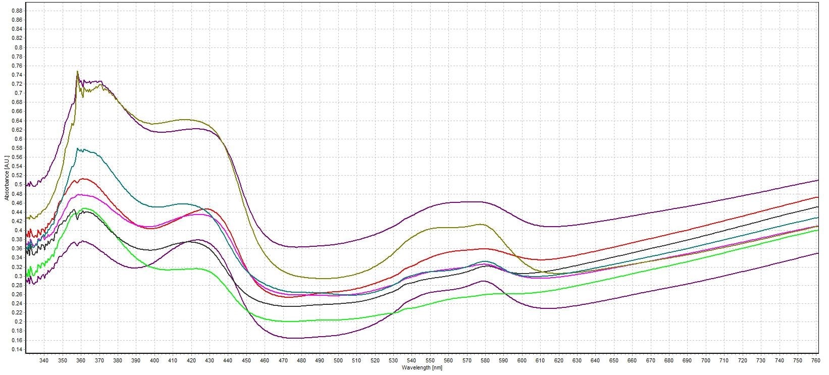
Figure 8: Volunteers with light hair and light-colored eyes
Conclusion:
In this experiment, we tested the absorbance and reflectance of skin samples to analyze skin pigmentation and protein structures. The skin pigmentation studied was melanin, and the protein studied was hemoglobin. Melanin is mainly located in the epidermis and is responsible for the pigmentation of the skin, hair, and eye color. Hemoglobin is located in the dermis which is below the epidermis. The skin reflectance measurements were used to show what colors were reflected from the skin samples and at what intensity. The more light the skin absorbed, the less light the skin then reflected.
Thirty volunteers offered to be a part of this experiment, and they were split into three spectral groups: dark hair and dark-colored eyes, dark hair and light-colored eyes, and light hair and light-colored eyes. The dark hair and dark-colored eye participants had the highest amount of melanin with the lowest skin reflectivity. They also had the least distinct M-shaped hemoglobin peak in the wavelength from 545-575 nm and the least uniform 420 nm hemoglobin hump. The dark hair and light-colored eyes had less melanin, but more defined hemoglobin peaks. The light hair and light-colored had the lowest amount of melanin and the highest skin reflectivity. They also had the most distinct M-shaped hemoglobin at 545-575 nm and the most uniform hemoglobin hump at 420 nm. This experiment showed the relationship between melanin, hemoglobin, reflectivity, and skin pigmentation with the help of an Avantes’ reflectance probe spectroscopic system.
Conducted by: Sara Beard (Chemistry Intern), Avantes Inc.
Theme: Skin Absorbance and Reflectance