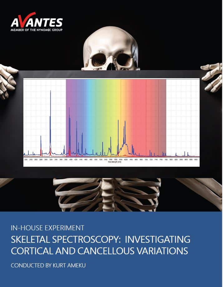
Skeletal Spectroscopy: Investigating Cortical and Cancellous Variations
 Introduction:
Along with trick-or-treating, carving pumpkins, and watching scary movies, skeletons are synonymous with the Halloween spirit. The inclusion of both skeletons and ghosts in Halloween festivities can be traced back to the Celtic festival of Samhain, from which our modern-day celebration of Halloween is derived. The holiday was believed to be a day where the boundary between the realm of the living and the realm of the dead became blurred, and the dead would return and walk amongst the living. With this “day of the dead” association, skeletons and bones in general became forever tied to Halloween. Generally, bones are divided into two categories: cortical and cancellous bone. Cortical bone, which makes up around 80% of skeletal muscle mass, is the rigid, outer layer that is responsible for a majority of weight and structural support along with storing fat in its bone marrow cavity. Cancellous bone is the spongy, inner layer of bone that is structured in a three-dimensional lattice known as trabeculae. This matrix structure arranges along stress lines to share some load bearing responsibilities, making it highly adaptable to changes in load and stress. Additionally, the space between the lattice in cancellous bone is filled with marrow and blood vessels. While these two types of bone vary significantly in structure, mechanical properties, and function, they are almost identical in terms of composition. Both types of bone are primarily composed of calcium phosphate, also known as hydroxyapatite, collagen, and water, along with some minor and trace elements. The main difference in composition between the two types of bone is calcium content, which is lower in cancellous bone. To determine calcium content, the amount of calcium must be compared to either the total mass of the sample or to the content of another present element that remains consistent. Often, a calcium-phosphorus ratio is used with bones since both elements are present in hydroxyapatite. One such technique to measure the presence of these and other elements in a sample is laser-induced breakdown spectroscopy, or LIBS.
In this LIBS-based experiment, our objective is to discern the distinct compositional disparities between cortical and cancellous bone. This technique requires the precise calibration of a laser, which, when focused on a bone sample, generates small plasma plumes. These plumes emit specific wavelengths corresponding to elements in the sample, revealing its composition. The bone sample used in this experiment (Figure 1) was cut multiple times to access both cortical and cancellous bone samples, both of which were measured using this spectroscopic technique, and individually scrutinized the resulting spectra. By matching measured peaks with the elements constituting bone composition, we directly compared the spectra to identify variations that could indicate whether a sample was cortical or cancellous bone, potentially through factors like the calcium-to-phosphorus ratio.
 My Cart